
News
Industry News > All the news
20 Dec 2025

Season's Greetings from eyesmart.com.au
19 Dec 2025

Safilo Acquires 25 Percent Stake in Inspecs Group
18 Dec 2025

Games and Cultural Sectors Get Vision-Impaired Accessibility Standards for XR
17 Dec 2025

New Zealand Study Finds Hearing and Vision Tests Drop in Aged Care
16 Dec 2025
Safilo Withdraws From Race to Acquire UK Eyewear Group Inspecs
15 Dec 2025

EssilorLuxottica Expands Med-Tech Footprint and Smart Eyewear Innovation
12 Dec 2025
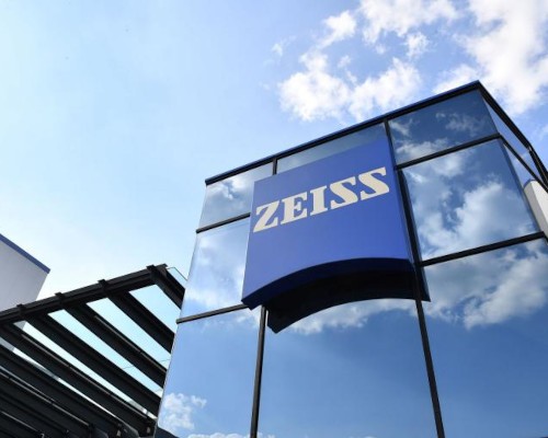
ZEISS Meditec CEO Steps Down Following Compliance Breach as Company Reports Solid Annual Growth
11 Dec 2025
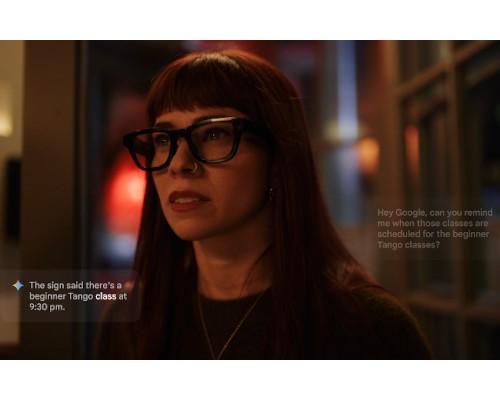
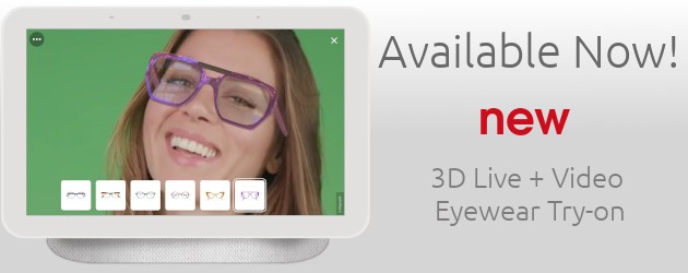
Google Returns to Smart Eyewear Market with AI-Powered Glasses in 2026
10 Dec 2025
ACO Opens Registrations for 2026 Certificate Courses, Introduces Advanced Ocular Therapeutics Program
09 Dec 2025
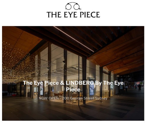
Lindberg Opens First Australian Boutique in Sydney CBD