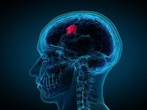
Eyecare Practitioners Are Well Placed To Diagnose Some Brain Tumours
 Despite being widely considered rare, brain tumours are responsible for more deaths of children and younger people than any other type of cancer (1).
Approximately 60% of brain tumour diagnoses comes from self-referral to Accident & Emergency (A&E). Which means there is a high percentage that have advanced disease at diagnosis (2). The most common age range for brain tumours in adults is 50 yrs and over and the risk increases with each decade, the peak age of presentation is between 60-79 yrs. (3).
Eyecare Practitioners (ECP) are a touchpoint for diagnosis as some brain tumours do have ocular presentations that prompt the visit. Approx. 28% of adults with a brain tumour report a visual impairment, (4) this increases up to 39% in children (5). Therefore it is imperative as primary care practitioners that all ECPs are able to detect subtle signs and symptoms of a brain tumour quite confidently. This equates to a quicker referral, accurate diagnosis and timely treatment.
Having a Optical Dispenser and/or a Practice Manager who is cognisant of some of these presenting signs & symptoms will help screen the importance of appointments to being symptom-led and triage them appropriately. What could initially start as a telephone conversation for a routine appointment may need to be escalated to a more urgent assessment or even a referral to a GP or even A&E.
Common risk factors for brain tumours include:
- Age
- Leukaemia
- Neurofibramatosis
- Previous episode of Lung and/or Breast Cancer
- Lifestyle- Smoking/Alcohol
- Family History of a brain tumour
Neurofibromatosis is a rare genetic disorder that causes typically benign tumours to form on nerve tissue. There are 3 different types, Type 1, 2 and 3. NF Type 1- these can develop anywhere in your nervous system including your brain, spinal cord and nerves. It is usually diagnosed in childhood or early adulthood. Affects approx. 1 in 3500 people worldwide. Lisch nodules on the Iris are an ocular characteristic (6).
These can be broken down into Non Vision Related Symptoms and Vision Related Symptoms.
Non Vision Related Symptoms:
- Headaches
- Seizures
- Vomiting
- Cognitive changes
Vision Related Symptoms are:
- Changes in Vision
- Newly Acquired Diplopia
- Anisocoria
- Papilloedema
- Nystagmus
Changes in Vision
Transient Visual Obscurations (TVOs) - tend to be reported as ‘dimming’ of the vision or ‘greying out’ of the vision which may be fleeting in duration lasting 1-3 secs. Patients find it difficult to isolate if it was unilateral or bilateral in nature (7). Sometimes people may not even mention it at all. However if it was suspicious in what was being described such as other symptoms that are listed in this article, it may be worthwhile going back and through very specific questions, to establish if TVOs have been experienced by the person.
Newly acquired Diplopia
This could be where the patient or the patients spouse describes this to you, face to face or over the phone that one of their eyes is turned in towards the their nose which would be a Cranial Nerve VI Palsy. The CN VI is the longest of the Cranial Nerves from its origin in the Pons to its point of attachment. Tumours can be insidious in developing and wrap themselves around the nerve itself restricting its movement leading to a horizontal diplopia which is worse for Distance than it is for reading. This needs urgent referral to A&E for Ophthalmological & Neurological investigation (8).
Pupil Involvement/Anisocoria
A Relative Afferent Pupillary Defect (RAPD) would not be present in the early stages of a brain tumour but may be present in the later stages as the Optic Nerve function is affected. An anisocoria is where there is a marked difference in the size of the pupils. One will be larger or smaller than the other. It may be long-standing in nature or it may be of a sudden onset. A sudden onset anisocoria noticed by the practitioner, family member, and/or partner should warrant further investigations. It may be more vasculopathic in origin such as a tear in the Internal Carotid Artery, but it should warrant immediate assessment.
Papilloedema
Papilloedema is a bilateral swelling of the Optic Discs due to high Intracranial Pressure, one cause of which is a brain tumour. It can be sometimes unilateral but that is quite rare.
Aetiology
An increase in Intracranial Pressure (ICP) within the brain. Approx. 80% of cases of ICP tend to be linked to a condition called Idiopathic Intracranial hypertension (IIH). This used to be referred as Benign IIH but its classification has changed due to its life threatening severity.
IIH is usually found in young woman of child bearing age with a slightly higher than normal BMI.
Other causes of raised ICP could be a Space Occupying Lesion (brain tumour), Venous Sinus Thrombosis (Blood Clot), or Meningitis.
Nystagmus
Usually Nystagmus comes under two different classifications either congenital or acquired.
This can be either a primary sign of a brain tumour or secondary Adverse Drug Reaction (ADRs) due to the medication prescribed after surgery. Seizures would tend to be a feature of brain tumour diagnosis journey in approx. 30% of cases (9). Anti-epilepsy or anti-convulsant medsication may be given post-surgery and one of the known side effects of theses medications that they may cause nystagmus (10).
Conclusion
There are certain ‘Red Flag’ signs and symptoms that ECPs need to be aware of in normal day to day practice that may require urgent referral to either another optometrist, GP or even A&E for suspect brain tumour. It is very difficult to list all the signs & symptoms of a brain Tumour but hopefully this article will better inform the ECP with specific symptoms that if heard on the shop floor, or heard on a telephone booking enquiry should be addressed immediately.
References:
- Cancer Research UK (2018), Brain, other CNS and intracranial tumours statistics
- NICE, Brain Tumours (primary) and brain mestases in adults NG99, 2018
- Kernick D et al Imaging patients with suspected brain tumours. Guidance for primary care. Br J Gen Pract 2008 (557) 880-885
- The Brain Tumour Charity, Losing Myself: The Reality of life with a brain tumour, 2015
- The Brain Tumour Charity: Losing my place: The reality of childhood with a brain tumour, 2015
- Cockey E et al, Neurofibromatosis type-1 associated brain tumours. Journal of rare diseases, research and treatment
- Seova N et al (2009) Papilloedema in patients with brain tumours, Neuro-Ophthalmology 33: 100-105
- Rucker, JC (2012) Diplopia, Third Nerve Palsies, and Sixth Nerve Palsies
- Liigant A et al (2001) Seizure disorders in patients with brain tumours Eur Neurol. 45:46-51
- Rowe F et al (2014) Interventions for eye movement disorders due to acquired brain injury (protocol) Cochrane Database of systematic Reviews, issue 9. DOI 10.1002/14651858
This article is based on an article for ABDO (Association of British Dispensing Opticians) by Lorcan Butler. He is a Dispensing Optician and am Optometrist with The Brain Tumour Charity in the UK. In his role as Optical Engagement Manager he is trying to educate the UK optical community of the importance of our sector as being a very important part of the referral process for further investigation for suspect brain tumours.


