


Researchers Develop Device For Early Diagnosis of Degenerative Eye Disorders
 Research into treatments to stop or limit the progression of degenerative eye disorders that can lead to blindness is moving ahead apace. But, at present, there is no device that can reliably diagnose these conditions before the first symptoms appear. These disorders, the best-known of which is age-related macular degeneration (AMD), involve changes to the eye’s photoreceptors. And they all have the same root cause: the deterioration of the retinal pigmentary epithelium (RPE), a layer of cells that sits behind the photoreceptors. The device developed at the Laboratory of Applied Photonics Devices (LAPD), École Polytechnique Fédérale de Lausanne (EPFL), Switzerland, observes changes in the RPE before the onset of symptoms, providing researchers with the first-ever in vivo images in which cells can be differentiated. Armed with this early detection capability, clinicians will be able to diagnose these disorders before irreversible symptoms occur. The results of the first clinical trial have been published in a paper in the journal Ophthalmology Science.
"The ability to observe morphological changes occurring in RPE cells is vital to the early detection of degenerative retinal disorders and to monitoring the efficacy of new treatments", says Laura Kowalczuk, a scientist at EPFL and at Jules-Gonin Eye Hospital.
In addition to causing AMD, the deterioration of the RPE is behind a number of other eye disorders, including retinitis pigmentosa and diabetic retinopathy. Located between the photoreceptors and the choroid (a thin layer of tissue containing the vessels that carry blood to the retina), the RPE plays an important role in maintaining visual function and preserving the health of the eye’s rods and cones. Several research groups have studied these cells under the microscope – in vitro – to determine their properties and to observe the morphological changes that occur with aging but also with the onset and progression of retinal disorders such as AMD and retinitis pigmentosa. Until now, however, there has been no simple and reliable method for observing the RPE in a live patient – in vivo – for early detection and ongoing monitoring of these conditions.
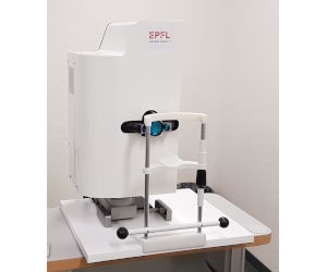
The researchers developed a clinical prototype in partnership with EarlySight, a spin-off from the same EPFL lab. With an exposure time of less than five seconds – a key speed advantage for potential diagnostic use – the camera is capable of capturing 100 raw images. Algorithms then align and aggregate the raw footage to produce a single, high-quality image on screen. The interface features five buttons, each corresponding to a predefined area of the eye, allowing the desired image to be selected. Users can also click anywhere on the diagram of the back of the eye to select the precise area they want to image.