


International Team of Researchers Makes Breakthrough in OCT Imaging
 An international team of researchers has achieved a significant technological breakthrough in the field of optical coherence tomography (OCT), a crucial form of light imaging. This breakthrough has the potential to revolutionize various applications in ophthalmology, dermatology, cardiology, and early cancer detection.
The research, led by experts from the University of Adelaide in Australia and the Technical University of Denmark (DTU), in collaboration with the Aerospace Corp in the USA and academics from the School of Physics and Astronomy at the University of St Andrews, has been published in the prestigious journal Science Advances.
Light imaging has witnessed remarkable advancements over the past decade, particularly in biomedical imaging, with its unparalleled ability to provide highly resolved image information with simplicity and ease of use. However, challenges still persist, particularly in recovering information from deep tissues. The scattering of light in tissue obstructs the retrieval of information at depth, similar to how light is scattered in a fog, making it difficult to see through.
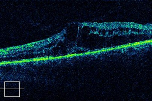
Optical coherence tomography (OCT) relies on the backscattering of light within the sample, occurring when light passes through different layers of cells, for instance. However, the scattering of light by cells and biological tissues poses a challenge for imaging. Obtaining a discernible signal beyond a depth of 1 mm is exceptionally challenging due to several factors, including signal interference from intervening tissue.
Conventionally, it has been believed that the OCT signal is predominantly affected by light that has undergone a single backscattering event, while light that is scattered multiple times hampers image formation. However, the research team discovered an alternative perspective: selectively collecting multiply scattered light can improve image contrast at depth, especially in highly scattering samples. They also demonstrated that this approach can be implemented easily with minimal additional optics by altering the light delivery and collection paths.
Gavrielle Untracht, the first author of the paper from DTU, expressed excitement about the study's results, stating, "The results of our study could be the start of a new way of thinking about OCT imaging. It's so exciting to contribute to such a technological breakthrough in the well-established OCT field!"
Professor Kishan Dholakia, from the School of Physics and Astronomy at the University of St Andrews, commented, "Our study breaks norms in optical imaging, and I believe it heralds a new path to recovering information at depth. OCT is an established method for obtaining useful information about human health, and our approach can enhance it even further."
Dr. Peter Andersen, co-corresponding author from DTU, added, "The unique configuration, supported by our modeling, should redefine our view on OCT signal formation – and we can now use this insight to extract more information and improve the diagnosis of diseases."
The team firmly believes that their breakthrough will challenge conventional methods and lead to a significant advancement in recovering images from deep tissues. Furthermore, they have both granted and filed intellectual property in this area, further boosting their confidence. The current OCT market was valued at US$1.3 billion in 2021 and is expected to triple by the end of the decade.
The research findings were published in the journal Science Advances.