

Portable AI Slit-Lamp Device Matches AS-OCT Accuracy in Anterior Chamber Screening
 A Japanese research team has developed a handheld, AI-integrated scanning slit-lamp device that delivers quantitative anterior-segment biometry at a fraction of the cost of conventional clinical equipment, a potential game-changer for community screening and teleophthalmology programs worldwide.
Published in Scientific Reports this March, the study from Tohoku University's Department of Ophthalmology describes a portable scanning slit-light device with a material cost below USD 500 that demonstrated strong agreement with anterior-segment optical coherence tomography (AS-OCT) for anterior chamber depth (ACD) measurement, the primary clinical output of interest.
The Problem It's Trying to Solve
The global burden of anterior-segment disease is well established. Conditions including primary angle-closure glaucoma, cataract, and keratoconus remain leading causes of avoidable blindness, with the burden disproportionately falling on low- and middle-income countries where access to specialist eye care can be up to four times lower than in high-income regions.
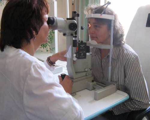
Even in well-resourced healthcare systems like Australia's, cost and access remain persistent barriers. Conventional slit-lamps cost several thousand dollars and are clinic-bound, while AS-OCT systems — the current quantitative gold standard for anterior-segment assessment — typically exceed USD 50,000. Neither is practical for community outreach, rural screening programs, or teleophthalmology deployments.
Existing portable and smartphone-based slit-lamp solutions have addressed the mobility problem to some degree, but the research team behind this study points out a critical gap: no commercially available slit-lamp, regardless of form factor, currently provides automated, quantitative anterior-segment measurement. All remain fundamentally qualitative and operator-dependent.
How the Device Works
The device combines portable slit-lamp imaging with on-device AI to generate anterior-segment measurements in a compact, self-contained unit. It captures a sequence of images across the eye and uses a lightweight model to identify anatomical structures and derive key metrics.
Designed for handheld use, the system incorporates adjustments to maintain consistency without the need for fixed positioning. All processing is performed locally, enabling use in remote or low-resource settings without reliance on internet connectivity.
Clinical Validation Results
In clinical testing, the device showed strong agreement with established imaging systems for measuring anterior chamber depth, with performance broadly in line with variability seen between existing clinical tools. This suggests it may be suitable for screening rather than diagnostic use.
While results were generally consistent, central corneal thickness measurements were less reliable due to current resolution limitations. The authors emphasise that further validation and refinement are needed before wider clinical adoption.
Segmentation Performance
The LWBNA-unet model, a lightweight U-Net architecture augmented with channel-attention modules, achieved training and validation Dice similarity coefficients of 0.936 and 0.947 respectively, with no evidence of overfitting. The model was trained on approximately 1,600 frames extracted from around 100 slit-scan videos, none of which overlapped with the AS-OCT validation cohort.
The model is specifically designed for deployment on resource-constrained hardware, including smartphones, Raspberry Pi units, and NVIDIA Jetson devices, a deliberate design choice to support global scaling without dependence on high-powered computing infrastructure.
Beyond ACD: A Multimodal Screening Platform
One aspect of the device that may attract particular interest from Australian practitioners is its multimodal capability from a single acquisition session.
Because the system captures full-colour visible-light images rather than the near-infrared grayscale of AS-OCT, it can visualise clinically important anterior-segment features including nuclear and cortical lens opacities, corneal irregularities characteristic of keratoconus, and peripheral anterior chamber configuration that are difficult or impossible to assess using infrared imaging alone.
In representative clinical cases presented in the paper, the device clearly differentiated a cataract case (ACD 2.71mm, iridocorneal angle 41.4°, lens density index 64.3) from a narrow-angle presentation (ACD 1.94mm, iridocorneal angle 15.1°, lens density index 34.9), with findings broadly consistent with paired AS-OCT data.
Beyond ACD and CCT, the research team indicates the same acquisition pipeline can support Van Herick ratio grading for angle-closure risk assessment, objective lens-opacity indices, dynamic pupillometry for autonomic function screening, and corneal curvature mapping for ectasia detection, functionalities that would traditionally require multiple dedicated instruments.
Limitations and the Road Ahead
The authors are candid about the current prototype's limitations. The validation cohort was modest in size and drawn from a single Japanese centre, meaning generalisability across ethnic groups, including Aboriginal and Torres Strait Islander populations with distinct ocular anatomy profiles, remains to be demonstrated. All participants were Japanese, and inter-ethnic differences in corneal diameter and anterior chamber configuration could influence device performance in broader screening programs.
Real-world deployment robustness under field conditions, including motion blur, poor patient cooperation, and challenging media opacities, will require dedicated prospective evaluation beyond the controlled clinical environment of this study.
The team identifies improved image resolution through reduced downsampling and more powerful edge-AI hardware as the primary pathway to bringing CCT accuracy to clinically actionable standards.
Future development plans include large-scale, multi-site dataset collection to train models capable of automated detection and grading of primary angle-closure disease, cataract, keratoconus, and other ectatic disorders, with outputs designed for integration into teleophthalmology dashboards.
Significance for Australian Eye Care
For Australian optometrists and ophthalmologists involved in community screening, rural outreach, or teleophthalmology services, the trajectory of this research is worth watching closely.
Australia's vast geography and chronically underserved rural and remote populations, where specialist ophthalmic access remains severely limited, represent exactly the use case this device is designed for. A sub-US$500 device capable of automated, quantitative anterior-segment screening without specialist operator training, running entirely offline on battery power, addresses a genuine gap in the current toolkit.
The device is not positioned as a replacement for AS-OCT or high-end slit-lamp examination in the clinic. Rather, the research frames it as a screening-grade tool capable of identifying patients who warrant further investigation, a role that could meaningfully extend the reach of anterior-segment disease detection into community and primary care settings.