


Routine Eye Scans Could Flag Heart and Brain Disease Risk Years Early
 A new UK study suggests the eye scans optometrists already perform every day could be repurposed as an early warning system for heart and brain disease potentially years before symptoms appear elsewhere in the body.
Researchers at the University of Manchester used artificial intelligence to comb through health data from more than 68,000 participants in the UK Biobank, a population-scale biomedical database. Using an AI tool the team built, called "Ret-AAE", they cross-referenced retinal imaging against genetic information and physiological measurements to map how strongly different eye features correspond to disease risk elsewhere in the body.
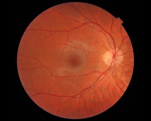
Two imaging types already standard in optometric and ophthalmic practice were assessed: optical coherence tomography (OCT), which produces 3D cross-sections of the retina, and colour fundus photography, the familiar flat retinal photograph used to monitor eye health. Both technologies are already in widespread use in practices.
The team found that the two scan types carry different but complementary signals about a patient's broader health. OCT findings tracked more closely with neurological traits, while colour fundus photographs were more strongly associated with cardiovascular indicators, pointing to a future where both scan types might be read together for a fuller systemic health picture.
On the genetic side, eye features identified by the AI were linked to genes involved in pathways associated with neurodegenerative conditions, including dementia. Separately, physiological analysis tied retinal features to blood pressure, blood vessel stiffness and heart function, reinforcing the long-suspected link between the retina's fine vascular network and cardiovascular health.
Lead author Dr Tom Julian, a Medical Research Council Clinical Research Training Fellow and eye doctor at the University of Manchester and Manchester Royal Eye Hospital, said the findings reinforced how much systemic information sits within retinal images, describing the eye as capable of revealing a broad picture of whole-body health that could help flag heart and brain disease risk ahead of time.
Co-author Dr Panos Sergouniotis, a Wellcome Clinician Scientist and honorary consultant at the University of Manchester, said while further work was still needed before such tests reach everyday clinical use, the long-term ambition is for routine eye examinations to double as a screening tool for broader disease prevention.
Professor Alejandro Frangi, who co-oversaw the work and holds a Royal Academy of Engineering chair, framed the implications squarely for the profession: using scans already available on high streets, an eye test may evolve into something well beyond a prescription check.
For practising optometrists, the research adds to a growing body of evidence positioning the practice chair as a potential frontline screening point for systemic disease leveraging equipment many practices already own, rather than requiring new capital investment. Whether and how such AI-derived risk scoring might eventually be integrated into clinical workflows, referral pathways or scope-of-practice frameworks remains to be worked through, including how it would translate to markets like Australia.
The study was published in Nature Cardiovascular Research on 16 June. It was funded by the Medical Research Council, Wellcome Trust, British Heart Foundation, the Royal Academy of Engineering, and the National Institute for Health and Care Research (NIHR) Manchester Biomedical Research Centre, among other supporters.
(photo credit: Topcon image by Julian)