


Lab-Grown Retinal Blood Vessel Cells Restore Sight-Threatening Damage in Mouse Trial
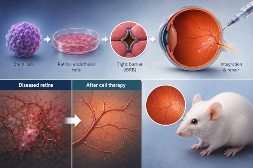 US researchers have developed a method to grow functional retinal endothelial cells from human stem cells, offering a potential new pathway to treating diabetic retinopathy and other retinal vascular diseases that remain poorly understood despite their status as leading causes of blindness.
The study, published in Nature Biomedical Engineering, was led by a team from Duke University's Department of Biomedical Engineering and represents one of the first reported methods for reliably generating retina-specific endothelial cells from human induced pluripotent stem cells (hiPSCs).
Addressing a gap in retinal research
The inner blood-retina barrier (iBRB), the specialised vascular structure that regulates what passes between blood vessels and retinal tissue, is central to maintaining eye health, but its breakdown is a hallmark of diabetic retinopathy and other microvascular diseases. Despite this, researchers have lacked a reliable, renewable source of human iBRB endothelial cells for study, as primary cells harvested from human tissue tend to lose their distinctive characteristics once removed from the body and cultured.
To solve this, the Duke team turned to hiPSCs, directing their differentiation into retinal endothelial cells (termed iRECs) by activating the Wnt–β-catenin signalling pathway specifically the Norrin–Frizzled4 axis known to be essential for retinal vascular development. The team also incorporated vitronectin and a compound called RepSox to support barrier maturation.
The resulting iRECs expressed key endothelial markers alongside retina-specific junctional proteins not typically found in generic endothelial cells, and showed genetic profiles that aligned more closely with primary human retinal endothelial cells than non-tissue-specific controls did.
Modelling disease and testing therapeutic potential
When exposed to high glucose and low oxygen conditions designed to mimic diabetic retinopathy, the iRECs showed a more pronounced disease response than standard endothelial cells including disrupted cell junctions and reduced barrier function suggesting they may offer a more clinically relevant model for studying the disease.
Perhaps most notably, when injected into the eyes of mice with oxygen-induced retinopathy, a widely used model for ischaemic retinal disease, the iRECs integrated with existing blood vessels, reduced pathological neovascularisation and vessel loss, and helped restore more normal vascular structure and reduced retinal permeability compared to controls.
The researchers also built functioning "iBRB-on-a-chip" microphysiological systems using the cells, and separately derived matching retinal pericytes from stem cells, demonstrating that the two cell types could self-organise into vascular networks resembling those found in the eye.
Implications for practice
While the work remains preclinical, the authors suggest the platform could eventually support both drug discovery and cell-based therapies for revascularising damaged retinas including the potential for patient-specific, isogenic cell lines. The senior author noted the approach may help address a long-standing barrier to progress in the field: the absence of biologically accurate, functionally active human retinal endothelial cells for research.
The team has filed a US provisional patent related to the derivation and therapeutic use of the cells. Further studies will be needed before any such approach could progress toward clinical application.