


Structured Light Could Turn a Little-Known Eye Reflex Into a New Retinal Health Test
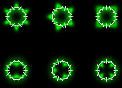 A quantum optics technique developed by Canadian and US researchers has made Boehm's brushes, a faint entoptic pattern most practitioners will never have heard of, visible enough to potentially underpin a future clinical test for retinal disease.
Most optometrists are familiar with Haidinger's brushes, the faint hourglass-shaped smudge some patients can see in central vision when looking at polarised light. Far fewer will know its peripheral cousin, Boehm's brushes: a dim, bowtie-shaped pattern that appears off-centre when a small polarised point source is viewed in the periphery, thought to arise from polarisation-sensitive scattering in the inner retina rather than macular pigment absorption.
Boehm's brushes have long been proposed as a potential biomarker of retinal integrity, on the theory that damaged retinal tissue would scatter polarised light differently to healthy tissue. The problem, according to a new study published in the Proceedings of the National Academy of Sciences, is that the phenomenon is usually too faint to reliably detect, even in people with healthy eyes, which has kept it out of clinical practice.
Turning two lobes into six
A research team led by the University at Buffalo and the University of Waterloo's School of Optometry and Vision Science has now shown that "structured light", beams engineered with spin-orbit coupling, combining circular polarisation with orbital angular momentum, can expand the classical two-lobed Boehm's brush pattern into a brighter, multilobed structure with up to six or more distinct lobes.
Rather than viewing a simple polarised point source, participants in the study looked at light shaped by a device called a Q-plate, which imprints a spatially varying polarisation pattern onto the beam. Depending on how the light was tuned, the number of visible lobes could be controlled, following a simple relationship between lobe count and the beam's topological structure.
Corresponding author Dusan Sarenac, now an assistant professor of physics at the University at Buffalo, said the engineered light changed the normally faint pattern into brighter patterns with a variable number of lobes, giving researchers several distinct ways to probe how a patient perceives the phenomenon and, by extension, retinal health.
What the testing involved
Eleven participants with healthy eyes screened with visual acuity charts, autorefraction, OCT and microperimetry to rule out macular abnormality viewed the structured stimulus through an annular aperture setup resembling a conventional eye exam. Six aperture sizes corresponded to retinal eccentricities between roughly 0.5° and 4° from fixation. A two-alternative forced-choice staircase procedure tracked the contrast needed for each participant to correctly identify the direction of rotation of the pattern.
Consistent with earlier classical measurements of Boehm's brushes, contrast detection thresholds fell steeply as the stimulus moved further from the fovea, levelling off at greater eccentricities an exponential decay pattern fitted with high consistency across all eleven participants. The pattern became reliably visible at around 1° of retinal eccentricity on average.
First author Dmitry Pushin, associate professor of physics at Waterloo, said that rather than simply asking participants whether they could see Boehm's brushes, the team measured how many lobes were seen, the contrast needed to detect them, and where in the visual field the pattern appeared.
Why it matters for clinical practice
The immediate application is not diagnostic. The current study only establishes a baseline in healthy eyes. But the authors frame the multilobed effect as a stepping stone toward screening tools for conditions such as early-stage age-related macular degeneration, diabetic maculopathy and glaucoma, where changes to retinal scattering structures or nerve fibre layer integrity could plausibly alter how patients perceive the pattern.
Sarenac's group is now planning to test the technique in patients with diagnosed retinal disease, to see whether damaged regions of the retina change the way the structured-light pattern is perceived, which would support its use as a functional biomarker alongside structural imaging such as OCT.
(Photo credit: Dusan Sarenac/University at Buffalo)