


New Lab-Grown Retina Model Rewrites Understanding of Vision Loss in Batten Disease
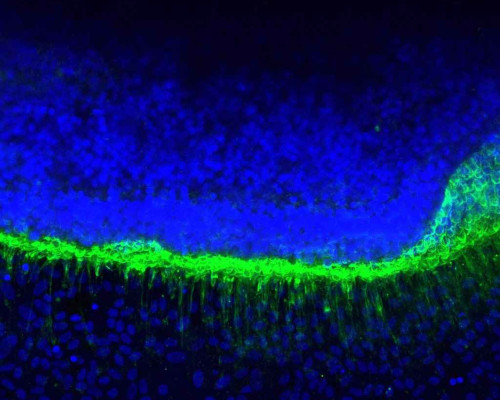 Findings suggest a support cell, not the photoreceptor itself, may be where treatment for the childhood condition should begin
A new 3D human retina model built by researchers in the United States has overturned a long-standing assumption about how vision loss begins in CLN3-Batten disease, a rare and fatal inherited condition that typically first appears in children as unexplained sight loss.
The study, published in Science Translational Medicine and led by the Flaum Eye Institute at the University of Rochester Medical Center in New York, points to a specific enzyme deficiency as an early driver of retinal damage and identifies a therapy already showing promise in preclinical testing.
A condition that starts in the eye
CLN3-Batten disease is a rare, inherited neurodegenerative disorder that most commonly presents in childhood. For many young patients, vision loss is the first noticeable symptom, often appearing years ahead of the seizures, cognitive decline and motor impairment that follow as the disease progresses. For clinicians working with paediatric patients, unexplained progressive vision loss combined with retinal changes has long been a red flag worth investigating further, even before a broader neurological picture emerges.
Until now, researchers have had limited tools for studying exactly how this early retinal damage unfolds. The affected tissue sits deep within the eye, making it difficult to observe the disease process directly in patients or replicate it faithfully in the lab.
Modelling the retina in a dish
To get around this, the Rochester team engineered a stem cell-derived 3D retina model that reproduces the interaction between two critical cell layers: the light-sensing photoreceptors and the retinal pigment epithelium (RPE), the support cells that sit beneath them and keep them functioning.
According to the research team, the model is the first to reliably reproduce the specific pattern of photoreceptor outer segment loss and degeneration seen in Batten disease patients, pathology that earlier models had struggled to capture with the same consistency.
Using the model, the researchers made a finding that challenges how the disease has traditionally been classified. CLN3-Batten disease has long been considered primarily a disorder of neurons, with photoreceptors assumed to be the first cells affected. The new data instead shows that dysfunction in the RPE support cells alone is enough to trigger photoreceptor degeneration suggesting the support cells, not the neurons, may be where the disease process actually begins in the eye.
Lead author Ruchira Singh, an associate professor at the Flaum Eye Institute, said the finding helps explain why sight loss tends to be one of the earliest symptoms families notice.
The lipid connection and a candidate therapy
Digging deeper into the mechanism, the team traced the RPE dysfunction to reduced activity of an enzyme called acid ceramidase, which normally helps regulate lipid balance inside cells. When acid ceramidase activity falls, lipids accumulate abnormally, contributing to the retinal damage observed in the model.
Armed with that target, the researchers trialled a recombinant form of the enzyme, known as rhAC, in both the lab-grown retina model and a large-animal model of the disease. The enzyme replacement therapy improved cellular health and reduced markers of retinal degeneration in both settings. The researchers noted that because the therapy restores enzyme function specifically in diseased cells without disrupting healthy ones, it stands out as a strong candidate for further development.
What it means for practice and the pipeline
There is currently no approved treatment that halts vision loss in CLN3-Batten disease, and several gene therapies and lipid-targeted drugs are already in various stages of development. The Rochester group suggests their findings could sharpen those efforts by highlighting the need to protect both the photoreceptors and the RPE layer supporting them, rather than focusing on neurons alone.
The retina model itself is likely to have a life beyond Batten disease. Because it recreates the photoreceptor–RPE interface with unusual fidelity, the researchers say it could also serve as a testing platform for other retinal diseases involving that same tissue interaction, a group that includes some more common causes of vision loss seen in optometric and ophthalmic practice.
The authors caution that rhAC therapy remains preclinical. Further work will need to establish long-term safety and efficacy before any trial in patients, and researchers are also examining whether the same treatment approach could address aspects of Batten disease beyond the eye.
The study drew on collaborators from the University of Rochester, the University of Tasmania, the Cleveland Clinic and the Icahn School of Medicine at Mount Sinai, and was funded by the US National Eye Institute, the ForeBatten Foundation and the Mangurian Foundation.